
 Mon - Sun 8:00 - 19:00
Many patients ask whether the procedure will cause significant discomfort. The definitive clinical reality is that placing an artificial titanium root into your jawbone involves absolutely zero sharp nociceptive signaling during the actual surgery. Because contemporary restorative protocols utilize highly targeted local anesthetics, you will only experience mild mechanical vibrations and localized pressure. The widespread apprehension surrounding this procedure typically stems from deep-seated psychological barriers and outdated historical dental experiences rather than current physiological realities. Once the targeted neural pathways are chemically blocked by modern pharmacological agents, the surgical site becomes completely desensitized. Furthermore, the alveolar bone tissue itself lacks the dense network of pain-sensing nerve fibers found within a natural tooth pulp, making the osteotomy phase surprisingly comfortable. Post-operative recovery generally mirrors the mild discomfort of a simple tooth extraction. This temporary inflammation is easily mitigated through standardized pharmacological regimens prescribed immediately after your session.
Clinical research on dental local anesthesia demonstrates that maintaining injection pressure below 306 mm Hg significantly reduces both nociceptive triggers and state anxiety among dental patients. Furthermore, utilizing advanced articaine-based buccal infiltrations achieves profound numbness in the mandibular molars with success rates ranging from 84% to 94%, bypassing older, more painful nerve block techniques. Finally, extensive studies utilizing visual analog scales confirm that minimizing the excess injected anesthetic volume directly yields a statistically significant reduction in post-operative edema and tissue trauma.
The Neurobiology of Dental Pain Transmission
Understanding the neurobiology of the oral cavity is crucial to alleviating procedural anxiety. The human jaw is innervated by the trigeminal nerve, which transmits sensory data to the brain. During surgery, local anesthetics temporarily disable these specific nerve branches. Consequently, while the brain remains conscious, the pathways responsible for transmitting pain signals are completely severed. This neurochemical blockade ensures that the surgical environment remains entirely painless.
To comprehend why the core question of do dental implants hurt is frequently misunderstood, one must examine the specific architecture of the peripheral nervous system. The oral cavity is a highly sensitive biomechanical environment. It relies on a vast network of sensory receptors designed to protect the airway and the digestive tract. These receptors transmit information through two primary types of nerve fibers. The A-delta fibers are heavily myelinated. They conduct electrical impulses at high velocities, resulting in the perception of sharp, acute, and highly localized pain. Conversely, the C fibers are unmyelinated. They transmit signals much more slowly, resulting in the perception of dull, aching, or throbbing pain that is difficult to pinpoint. When a biological tooth becomes infected, the inflammation primarily stimulates the C fibers within the dental pulp, causing excruciating, radiating agony.
However, the anatomical landscape shifts dramatically when discussing the jawbone itself. The alveolar bone, which serves as the foundational structure for the artificial titanium root, possesses a radically different neurological profile compared to a living tooth. The internal matrix of the bone is remarkably sparse in nociceptors (pain-sensing nerve endings). The majority of the sensory innervation is located in the soft gingival tissues and the periosteum, which is the thin, fibrous membrane enveloping the outer surface of the bone. Therefore, once the clinician successfully desensitizes the superficial gum tissues and the periosteum, the underlying bone can be manipulated, drilled, and sculpted without triggering any nociceptive response. The structural reality is that bone tissue simply does not have the biological hardware to register sharp surgical pain.
This anatomical advantage is further leveraged by precise surgical mapping. Before any physical intervention occurs, modern dental professionals utilize Cone Beam Computed Tomography (CBCT) to create a three-dimensional map of the patient’s craniofacial anatomy. This digital blueprint allows the surgeon to identify the exact spatial coordinates of major nerve bundles, such as the inferior alveolar nerve in the mandible and the sinus cavities in the maxilla. By maintaining a strict safety margin of several millimeters away from these critical neural pathways, the surgical site remains confined to the neurologically barren regions of the alveolar ridge. The intersection of this anatomical reality with advanced imaging technology completely neutralizes the biological potential for intraoperative pain.
Modern Dental Anesthesia Mechanisms
Modern pharmacological agents work by targeting the cellular membranes of nerve fibers. Anesthetics reversibly bind to sodium channels, preventing the electrical impulses required for pain perception from ever forming. By halting this cellular depolarization, nociceptive impulses associated with surgical stimuli cannot reach the central nervous system. This highly localized chemical intervention is what allows clinicians to perform complex rehabilitations without the patient feeling anything.
The administration of local anesthesia is an exercise in applied biochemistry. The human nervous system communicates via action potentials, which are rapid changes in the electrical charge across a cell membrane. At rest, a nerve cell maintains a negative internal charge. When a pain stimulus occurs, microscopic protein channels in the membrane open, allowing positively charged sodium ions to flood into the cell. This sudden influx of positive charge triggers an electrical impulse that travels up the nerve fiber to the brain, registering as pain. Local anesthetics, such as Lidocaine, Articaine, and Bupivacaine, act as chemical roadblocks. Their molecular structure allows them to physically enter the sodium channels and plug them from the inside. Without the influx of sodium, the nerve fiber cannot fire. The brain simply never receives the message that a surgical procedure is occurring.
The efficacy of these chemical agents is dictated by their lipid solubility, protein binding capacity, and vasodilatory properties. A highly lipid-soluble anesthetic can easily penetrate the fatty myelin sheath surrounding the nerve, ensuring a faster onset of profound numbness.
| Pharmacological Agent | Lipid Solubility | Protein Binding | Clinical Duration | Primary Application in Implantology |
| Lidocaine 2% | Moderate | 64% | 60 – 90 minutes | Standard mucosal infiltrations and short restorative procedures. |
| Articaine 4% | Very High | 95% | 60 – 120 minutes | Dense mandibular bone infiltrations due to superior tissue diffusion. |
| Bupivacaine 0.5% | High | 95% | 240 – 480+ minutes | Extensive full-arch surgeries and prolonged post-operative pain control. |
The introduction of Articaine into the dental armamentarium represents a monumental shift in patient comfort. Historically, numbing the lower jaw required an Inferior Alveolar Nerve Block (IANB), a technique that necessitates deep needle penetration and carries a higher risk of temporary nerve parasthesia. The IANB is statistically rated by patients as the most uncomfortable injection due to the depth of tissue penetration required. Articaine, however, possesses a thiophene ring in its molecular structure, making it exceptionally lipid-soluble. This unique characteristic allows it to diffuse directly through the dense cortical bone of the mandible. Consequently, clinicians can now achieve profound pulpal and soft tissue anesthesia in the lower jaw using a simple, shallow buccal infiltration. Clinical trials confirm that this less invasive technique yields success rates between 84% and 94%, completely negating the need for the deeper, more intimidating nerve block.
To prolong the duration of the numbing effect, these anesthetic solutions are formulated with a microscopic concentration of a vasoconstrictor, typically Epinephrine (Adrenaline). By constricting the local blood vessels, the epinephrine prevents the systemic bloodstream from rapidly washing the anesthetic molecules away from the surgical site. This ensures that the patient remains entirely comfortable throughout even the most complex, multi-hour full mouth rehabilitations. However, the precise calibration of these vasoconstrictors is paramount. Patients presenting with systemic cardiovascular diseases require highly customized formulations to prevent adverse hemodynamic responses, demanding a sophisticated level of pharmacological mastery from the surgical team.
Is Dental Implant Surgery Painful? The Myth vs. Clinical Reality
People frequently ask is dental implant surgery painful when comparing it to historical dental trauma. The reality is that patients often confuse harmless mechanical vibration with actual tissue injury. While you will feel pressure and hear the clinical instruments, true pain is physiologically impossible under proper anesthesia. Distinguishing between tactile proprioception and nociception allows patients to remain calm and comfortable throughout the restorative appointment.
To fully understand the patient experience, we must dissect the phenomenon of proprioception. Proprioception is the body’s ability to sense its position, movement, and the mechanical forces acting upon it. This sensory data is gathered by specialized mechanoreceptors located within the periodontal ligaments, the temporomandibular joint, and the surrounding facial musculature. Unlike nociceptors, which are completely silenced by local anesthetics, some mechanoreceptors remain partially active. Their biological imperative is to prevent the jaw from hyperextending and to regulate bite force.
During the osteotomy phase, the surgeon utilizes specialized implant drills operating at highly calibrated torques and speeds (typically between 800 to 1200 RPM with copious saline irrigation) to precisely prepare the bone bed. As the drill gently shapes the alveolar ridge, it generates deep, low-frequency vibrations. These vibrations resonate through the dense skeletal architecture of the skull, conducting sound directly to the inner ear via bone conduction. For a patient lying in the surgical chair, this sensory input is profound. The hum of the motor and the deep physical pressure are undeniable.
However, the critical cognitive leap required for patient comfort is recognizing that vibration does not equal tissue damage. The human brain, particularly when primed by preoperative anxiety, is biologically programmed to interpret loud noises and deep pressure near the cranium as an immediate physical threat. This cognitive misinterpretation triggers a sympathetic nervous system response, causing the patient to instinctively tense their muscles and brace for an impact that will never arrive. The perceived discomfort is an illusion synthesized by the brain’s defense mechanisms. The actual surgical site remains entirely numb and biologically insulated from nociceptive trauma. By educating patients on the physiological distinction between harmless mechanical pressure and actual pain, clinicians can dramatically reduce intraoperative anxiety and facilitate a relaxed, cooperative surgical environment.
Advanced Injection Techniques: Eradicating the Needle Fear
Before we delve into the mechanical mastery of pain control, visual evidence of modern equipment provides immediate psychological reassurance.
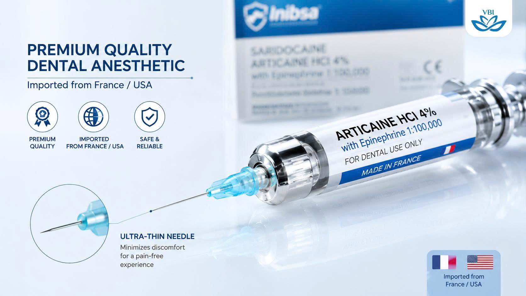
The initial administration of numbing agents is often the most feared aspect of the visit. However, discomfort does not come from the ultra-thin needle, but rather the rapid expansion of gingival tissues. By utilizing computerized delivery systems and maintaining an injection pressure below 306 mm Hg, clinicians ensure a gentle, unnoticeable fluid release. This biomechanical mastery completely transforms the patient’s sensory experience during the initial phase.
The widespread phobia surrounding dental injections is largely based on outdated, manual administration techniques. When a practitioner manually depresses the plunger of a traditional anesthetic syringe, human error often results in inconsistent fluid pressure. If the anesthetic solution is forced into the tightly bound mucoperiosteum too rapidly, it causes microscopic tissue tearing and massive cellular displacement. This physical trauma, rather than the penetration of the needle itself, is the primary source of injection pain.
Modern dental facilities utilize advanced fluid dynamics to eliminate this issue. Clinical studies utilizing invasive sphygmomanometers demonstrate a direct, undeniable correlation between injection pressure and perceived pain. To neutralize this, elite practitioners employ computerized local anesthesia delivery systems (C-CLAD). These microprocessor-controlled devices regulate the flow rate of the anesthetic fluid with absolute precision. By constantly measuring tissue resistance and ensuring the injection pressure remains strictly below the pain-triggering threshold of 306 mm Hg, the device facilitates a slow, highly controlled diffusion of the anesthetic. This technology creates an “anesthetic pathway,” essentially numbing the tissue micro-millimeters ahead of the advancing needle. The resulting experience is so gentle that many patients are unaware the injection has even occurred.
Furthermore, clinicians utilize the Gate Control Theory of Pain to their advantage. This neurological theory posits that non-painful tactile inputs can close the “nerve gates” to painful inputs, preventing nociceptive signals from traveling to the central nervous system. A prime example is the application of upper lip compression during maxillary infiltrations. By applying firm, steady pressure or a rapid vibratory stimulus to the lip and surrounding mucosa just prior to and during the injection, the mechanoreceptors are flooded with data. This overwhelming tactile signal outcompetes the minor discomfort of the needle penetration, effectively distracting the brain and significantly reducing the perception of pain.
Topographical anatomy dictates the specific approach. Maxillary infiltrations are generally straightforward due to the highly porous nature of the upper jawbone. Palatal injections, historically dreaded due to the dense, tightly adhered tissue on the roof of the mouth, are now managed seamlessly. Clinicians use profound topical anesthetics containing 20% Benzocaine, combined with pressure techniques (using a mirror handle to apply intense pressure next to the injection site), to completely mask the fluid release. For the mandible, the transition from traumatic nerve blocks to gentle Articaine infiltrations represents the zenith of patient-centric care.
Managing Pain After Dental Implant Surgery
Visualizing the standard post-operative care package illustrates the meticulous preparation involved in modern dental tourism recovery.
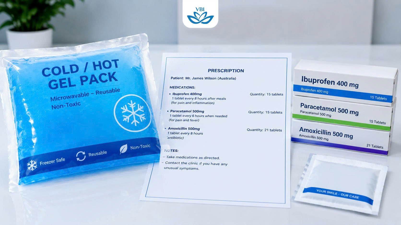
Addressing pain after dental implant surgery requires a proactive pharmacological strategy. Once the clinical numbing wears off, the body initiates an inflammatory cascade to heal the osteotomy site. This natural swelling is effectively suppressed using non-steroidal anti-inflammatory drugs taken preemptively. When patients strictly follow the prescribed medication and cryotherapy protocols, post-operative discomfort remains minimal, peaking at 48 hours before rapidly subsiding.
The cessation of the local anesthetic triggers the commencement of the biological healing phase. Within two to four hours post-surgery, as the sodium channels unblock and normal nerve function resumes, the immune system initiates an inflammatory response. This is not a complication; it is a vital biological necessity. To facilitate tissue repair and bone integration, the body releases a surge of chemical mediators, most notably prostaglandins and leukotrienes. These lipids act as cellular distress signals, causing massive vasodilation. The resulting influx of blood plasma, white blood cells, and restorative nutrients into the surgical site causes the gingival tissues to swell. This edema exerts pressure on the surrounding nerve endings, translating into the dull, throbbing sensation typical of post-operative recovery.
Managing this inflammatory curve is a science of timing. The cardinal rule of pain management is preemption. Clinicians prescribe Non-Steroidal Anti-Inflammatory Drugs (NSAIDs), such as Ibuprofen or Meloxicam, to be taken before the local anesthesia completely dissipates. NSAIDs function by inhibiting the Cyclooxygenase (COX-1 and COX-2) enzymes. By blocking these specific enzymes, the medications halt the synthesis of prostaglandins at the source. This chemical blockade prevents the pain signal from ever reaching peak intensity.
| Recovery Timeline | Biological Process | Recommended Pain Management Protocol |
| Hours 1 – 4 | Local anesthesia gradually metabolizes. Nerve function returns to baseline. | Consume the first dose of prescribed NSAIDs before numbness entirely fades. |
| Hours 4 – 24 | Prostaglandin release triggers initial vasodilation and localized tissue edema. | Apply cryotherapy (ice packs) externally: 15 minutes on, 15 minutes off. |
| Hours 24 – 48 | Inflammatory cascade reaches its absolute peak. Maximum swelling occurs. | Continue NSAIDs. Switch to warm compresses to promote lymphatic drainage. |
| Days 3 – 7 | Edema rapidly subsides. Cellular repair and early osseointegration begin. | Taper off NSAIDs. Manage residual soreness with mild analgesics like Paracetamol. |
A critical, yet often overlooked variable in post-operative comfort is the total volume of anesthetic fluid injected during the procedure. Clinical data reveals a direct correlation between excessive local anesthetic volume and increased post-operative morbidity. Over-saturating the surgical site with fluid causes severe tissue distension, essentially stretching the gingival fibers beyond their physiological limits. This unnecessary trauma dramatically exacerbates post-operative swelling and extends the pain curve. Elite practitioners mitigate this by utilizing high-concentration anesthetics delivered with pinpoint accuracy, minimizing the fluid burden on the soft tissues and ensuring a vastly superior recovery experience. For a comprehensive understanding of the entire therapeutic journey, review your complete surgery timeline.
Surgical Variables Affecting the Healing Timeline
The surgical methodology employed directly dictates the severity of the post-operative inflammatory response. The evolution of digital dentistry has ushered in an era of minimally invasive protocols that preserve critical blood supply and minimize soft tissue trauma. Consequently, the healing timeline is drastically compressed, allowing international patients to resume their holiday itineraries with minimal interruption.
Historically, implantology relied on extensive mucoperiosteal flaps. The surgeon would make a broad incision along the crest of the ridge and physically peel back the gum tissue to expose the underlying bone. This gross elevation severs the delicate periosteal blood vessels, triggering a massive inflammatory response and significant post-operative bruising. While this technique remains necessary for complex cases requiring extensive bone grafting, it is no longer the default standard of care for straightforward placements.
The contemporary gold standard is flapless, guided implant surgery. Utilizing the digital blueprint generated by the CBCT scan, a highly precise surgical guide is 3D printed. This resin template fits seamlessly over the patient’s existing dentition. It features titanium sleeves that dictate the exact angulation, depth, and trajectory of the surgical drills. Because the precise topography of the bone is known, the surgeon simply uses a tissue punch to remove a tiny circular core of gingiva, exactly the diameter of the implant. The entire osteotomy is performed through this microscopic aperture. The periosteum remains entirely undisturbed, preserving the crucial micro-vascular network. The result is a near bloodless procedure with virtually zero post-operative swelling and a negligible pain curve.
The density of the jawbone also plays a pivotal role in the surgical experience. Bone is categorized into four distinct densities, ranging from D1 (dense, oak-like cortical bone, typically found in the anterior mandible) to D4 (soft, styrofoam-like trabecular bone, often located in the posterior maxilla). Drilling into D1 bone requires higher torque, generating more friction and more pronounced intraoperative vibrations. Conversely, D4 bone requires minimal preparation; the site is often expanded using gentle hand instruments called osteotomes rather than high-speed drills. While the D4 approach involves less vibration, it necessitates a longer healing period for osseointegration to finalize. A thorough preoperative analysis ensures the patient’s expectations regarding surgical sensations and recovery timelines are perfectly aligned with their unique biological reality.
Age, Anatomy, and Cardiovascular Considerations
Older demographics seeking full arch restorations possess unique anatomical characteristics that influence surgical comfort. As natural alveolar bone resorbs over time, the overlying palatal mucosa loses its tautness. This biological shift means less mechanical pressure is required to administer local anesthetics, resulting in a significantly less painful injection. However, clinicians must carefully balance anesthetic efficacy with cardiovascular constraints regarding vasoconstrictor usage in older patients.
The aging process inherently alters the topography of the oral cavity. Following the loss of natural dentition, the alveolar ridge undergoes a continuous process of disuse atrophy. Without the mechanical stimulation previously provided by the tooth roots during mastication, the osteoclasts (cells responsible for bone resorption) outpace the osteoblasts (cells responsible for bone formation). Over decades, this results in severe vertical and horizontal bone loss.
While this skeletal degradation presents profound challenges for implant placement—often requiring advanced angled techniques like the All-on-4 protocol—it inadvertently provides a localized benefit regarding anesthesia administration. In younger adults, the mucoperiosteum on the palate is exceptionally taut and firmly bound to the underlying bone. Injecting fluid beneath this unyielding membrane requires significant mechanical pressure, which translates into an acute, burning pain. However, in older adults suffering from severe bone resorption, this palatal mucosa becomes loose and flaccid. The anesthetic fluid easily flows into the sub-epithelial space with minimal resistance. Consequently, the administration of local anesthesia for older patients is clinically proven to be administered with far less force, causing significantly less pain.
Despite this anatomical advantage, managing the older patient demographic requires extreme pharmacological vigilance. A substantial portion of patients undergoing full-arch rehabilitations present with complex medical histories, including hypertension, ischemic heart disease, or past myocardial infarctions. These systemic realities severely limit the use of Epinephrine (adrenaline) in the anesthetic solution. High doses of vasoconstrictors can precipitate tachycardia or dangerous spikes in blood pressure. The surgical team must utilize specialized anesthetic formulations, such as Mepivacaine 3% plain (which contains no vasoconstrictor), or utilize advanced ultrasound-guided nerve blocks to achieve profound numbness without risking cardiovascular instability. The intersection of geriatric anatomy and internal medicine demands an elite level of interdisciplinary care.
The Psychological Dimension: Anxiety-Induced Hyperalgesia
Psychological distress acts as a potent amplifier for physical sensation. Anxiety triggers the release of stress hormones, drastically lowering a patient’s natural pain threshold through a process called hyperalgesia. In this hyper-vigilant state, minor tactile pressures are misinterpreted by the brain as severe threats. Managing this cognitive response through deep sedation, pristine clinical environments, and continuous verbal reassurance is critical for total patient comfort.
The mind-body connection in oral surgery is not a theoretical concept; it is an undeniable physiological reality. When a patient enters the operatory harboring severe dental phobia, their sympathetic nervous system is heavily activated. The adrenal glands dump massive quantities of cortisol and epinephrine into the bloodstream. This neurochemical cascade puts the entire central nervous system on high alert. The brain begins aggressively scanning the environment for threats. In this state of heightened arousal, the pain threshold plummets. A phenomenon known as hyperalgesia occurs, where a normally innocuous stimulus—such as the cold touch of a dental mirror or a drop of water on the tongue—is instantly interpreted as sharp, agonizing pain.
This psychological tension creates a dangerous feedback loop. Fear induces muscle rigidity. When a patient involuntarily tenses their facial musculature and stiffens their neck, it significantly increases the localized resistance during the anesthetic injection, thereby creating the very pain they were attempting to avoid. Furthermore, anxiety accelerates the patient’s basal metabolic rate. This rapid metabolism causes the body to process and eliminate the local anesthetic much faster than normal, leading to sudden, unexpected breakthroughs in sensation during the procedure.
To disrupt this cycle, comprehensive clinics deploy robust behavioral and pharmacological interventions. In cases involving extensive rehabilitations or profound clinical phobia, conscious sedation protocols are strongly recommended. Intravenous (IV) sedation utilizing agents like Midazolam or Propofol induces a state of profound relaxation and anterograde amnesia. The patient remains capable of maintaining their own airway and responding to basic verbal commands, but they are entirely detached from the surgical environment.
It is crucial to note that sedation does not replace local anesthesia; it supplements it. The sedatives alter the patient’s state of consciousness, while the local anesthetics remain exclusively responsible for blocking the peripheral nociceptive signals at the jawbone. For surgeries with an average duration of 90 minutes or more, ensuring the local anesthetic remains robustly effective is paramount, as the wearing-off of the numbing agent will immediately disrupt the sedated state and cause patient distress. For an academic review of how these distinct mechanisms synergize, explore the latest literature on modern dental anesthesia techniques.
The Economic and Psychological Relief of Dental Tourism
Financial toxicity represents a massive psychological burden for patients seeking full mouth rehabilitations in Western countries. Traveling to specialized clinics in Southeast Asia drastically reduces this economic stress, which inversely lowers physical tension in the surgical chair. Knowing you are receiving world-class care at a fraction of the domestic cost provides profound psychological relief. This holistic peace of mind directly contributes to a smoother healing process.
The fear of pain is invariably intertwined with the fear of financial ruin. Patients traveling from Australia, New Zealand, the United Kingdom, or the United States frequently face extreme economic anxiety regarding restorative dentistry. The sheer cost of replacing missing teeth in Western nations—often exceeding tens of thousands of dollars for a single arch—creates an immense psychological burden. This phenomenon, clinically recognized as financial toxicity, demonstrably lowers the overall quality of life and severely exacerbates preoperative anxiety. When a patient sits in a surgical chair knowing the procedure is depleting their life savings, their physiological stress markers are dangerously elevated.
By accessing elite healthcare in regions like Hanoi, patients bypass the inflated costs associated with excessive domestic operational overhead, bloated administrative staff, and exorbitant defensive medical malpractice insurance premiums. The realization that they are receiving identical, if not superior, clinical standards provides profound psychological relief. These facilities utilize authentic, globally recognized implant brands (such as Straumann and Nobel Biocare) manufactured from Titanium Grade 5. They employ advanced digital workflow systems and strictly adhere to rigorous global infection control protocols, including the Association for the Advancement of Medical Instrumentation (AAMI) standards for autoclave sterilization.
This economic relief acts as an incredibly potent form of preoperative sedation. When the financial trauma is removed from the equation, the patient’s baseline stress drops precipitously. This directly correlates to lower muscle tension, a more cooperative posture in the surgical chair, and a vastly reduced risk of procedural trauma. The synergy between unparalleled financial peace of mind, meticulous surgical precision, and advanced pharmacological pain management creates a holistic healing environment. The journey transforms from a terrifying medical obligation into an empowering, life-changing restoration of health and confidence.


Bài viết liên quan
Dental Implant Failure Symptoms: Causes & How to Fix It
Dental implants represent a highly predictable restorative solution in modern dentistry, but they are not...
The Hidden Risks of Cheap Dental Implants Overseas
Seeking affordable dental care abroad sounds highly appealing. But what are the actual cheap dental...
Using Super for Dental Implants: Australian Guidelines 2026
Many patients ask about using super for dental implants and navigating the ATO compassionate release...